Engineered and patient DMD agree descriptively
GSE233606 provides an unselected gene-axis bridge between engineered DMD and a patient line. It is descriptive, not donor-level inference.
Measured DMD evidence · Stage C2
This release adds biological-repeat-aware Reactome tests for GSE272233 and sample-level external disease validation. It strengthens disease-context evidence and provides reference axes for a prespecified prospective candidate-validation study.
Evidence chain
GSE233606 provides an unselected gene-axis bridge between engineered DMD and a patient line. It is descriptive, not donor-level inference.
PRJNA772047 uses five reported donor/sample pseudobulks. PRJNA1218493 and mouse GSE156497 remain directional sensitivity layers.
GSE272233 uses three reported biological repeats per group. No dual-supported pathway reverses across all three mutation backgrounds.
Organoid evidence is shown as a heterogeneity layer and is not pooled into a universal DMD response.
NAR main figure
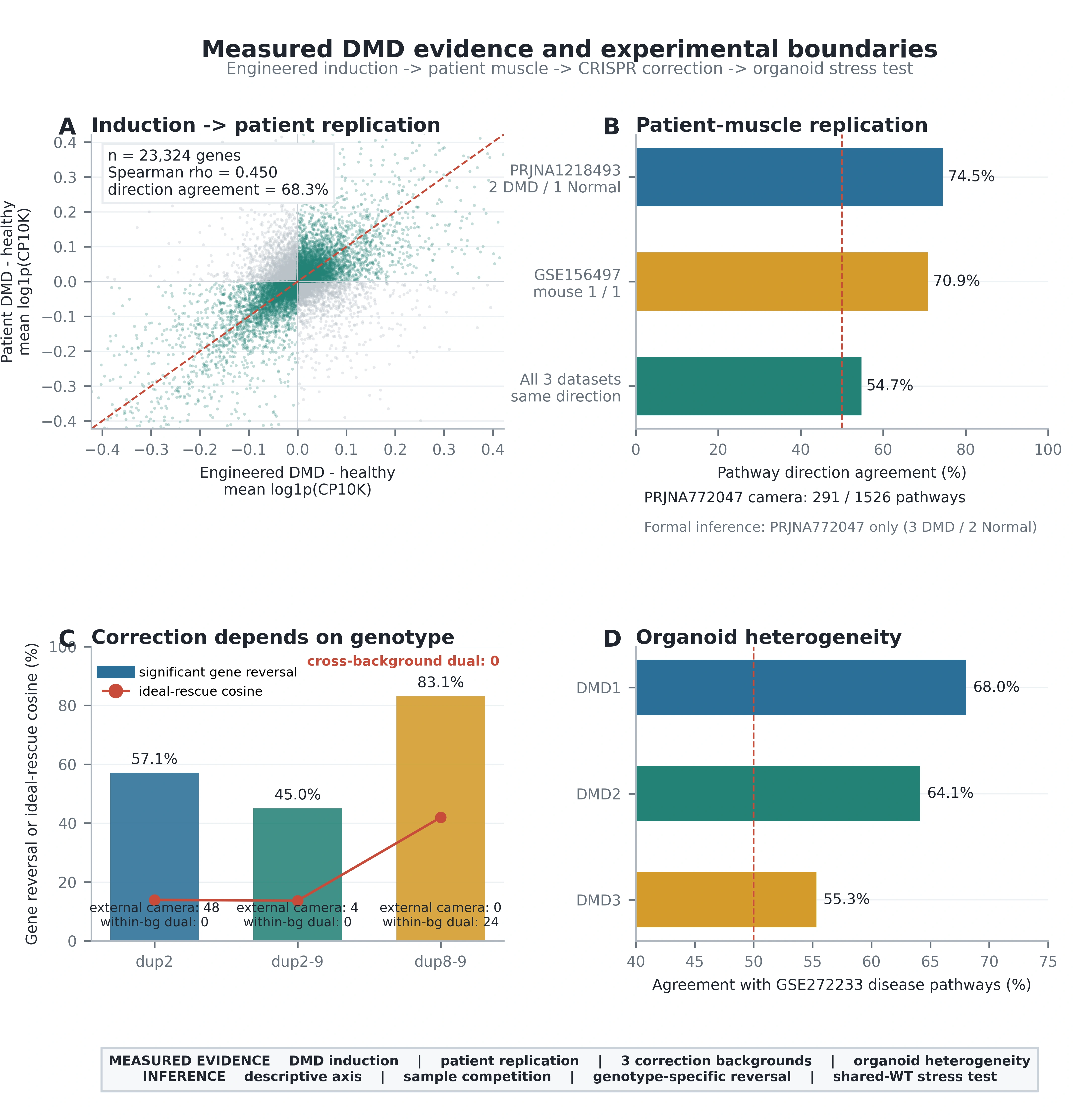
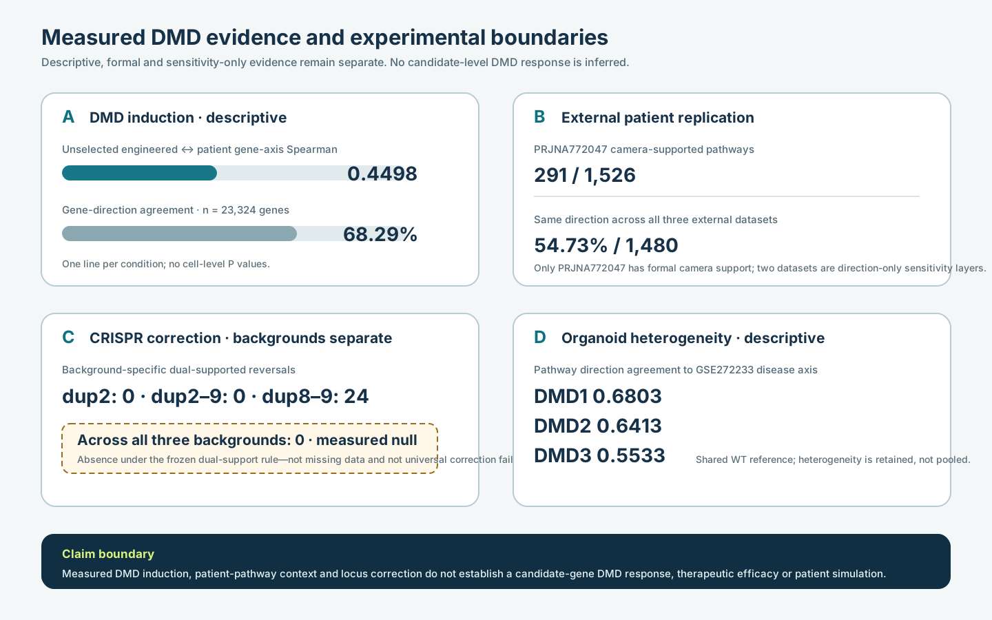
Long description. Panel A shows a positive descriptive association between the unselected engineered-DMD and patient-DMD gene axes (Spearman 0.4498 across 23,324 genes; direction agreement 68.29%). Panel B separates one formal patient pseudobulk test from two directional sensitivity datasets: 291 PRJNA772047 pathways have camera support, while 54.73% of 1,480 shared pathways keep the same direction across all three external datasets. Panel C compares three DMD-locus correction backgrounds; 24 dual-supported pathway reversals occur only in dup8–9 and none are shared by all backgrounds. Panel D shows decreasing organoid-to-disease pathway agreement across DMD1, DMD2 and DMD3 (0.6803, 0.6413 and 0.5533), preserving heterogeneity rather than pooling it away.
| Panel | Evidence layer | Metric | Value | n | Inference |
|---|---|---|---|---|---|
| A | DMD induction | Unselected gene-axis Spearman | 0.4498 | 23,324 genes | Descriptive; one line per condition |
| A | DMD induction | Direction agreement | 68.29% | 23,324 genes | Descriptive; no cell-level P values |
| B | Patient replication | camera-supported pathways | 291 | 1,526 pathways | 3 DMD vs 2 normal donors/samples |
| B | External sensitivity | Same direction in all three datasets | 54.73% | 1,480 pathways | Directional agreement only |
| C | CRISPR correction | Background-specific dual reversals | 24 | 1,559 pathways | Observed only in dup8–9 |
| C | Cross-background correction | Dual reversal in all backgrounds | 0 · measured null | 3 backgrounds | Measured absence under the frozen rule |
| D | Organoid heterogeneity | Direction agreement range | 0.5533–0.6803 | 1,511 pathways | Descriptive; shared WT reference |
Methods boundary. Panel A is descriptive. Panel B uses camera only for PRJNA772047 and direction-only sensitivity for the other datasets. Panel C keeps mutation backgrounds separate and uses the frozen dual-support rule. Panel D is a heterogeneity display, not a pooled universal response. Open figure-generation code.
Primary statistical readout
The 492 fixed-effect cross-background reversals are explicitly exploratory because WT is non-isogenic and heterogeneity is substantial. They are not promoted to validated virtual-cell predictions.
Reusable objects
Independent rebuild receipt
The path-isolated rebuild regenerated GSE233606 induction, GSE272233 correction and camera tests, three external pseudobulk pathway layers, GSE277637 heterogeneity and Figure 8. Numeric tables and metadata-independent figure pixels match the released analysis.
Boundary: this is path/process-isolated computational reproduction using the same system Python/R packages, not a container-level independent-host reproduction and not biological validation.
Supported: Measured DMD-locus induction/correction and sample-level external disease pathway replication with explicit small-n limitations.
Not supported: Candidate-gene response prediction in DMD cells, therapeutic efficacy, patient-level simulation and clinical decision support.